Welcome to a special year-end Brain Bits! Today I’ll discuss some of my favorite brain-related research from 2017.
*Note: “Best of” = random papers that I liked. Apologies to the authors of all the other awesome 2017 papers that are not mentioned here!
How the brain creates an internal compass
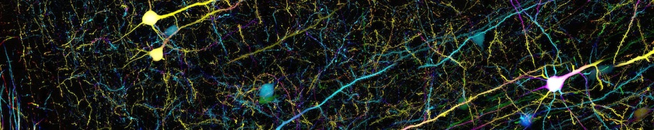
My favorite neuroscience story of the year was the discovery of the mechanism that creates an internal compass in the brain. Back in 2015, Vivek Jayaraman’s lab at HHMI’s Janelia Research Campus discovered that fruit flies contain an internal compass that tells them which way they’re going as they wander around the world (as I described in my May 2015 Brain Bits). This compass is contained within a doughnut-shaped brain structure called the ellipsoid body. At any given moment, just one wedge of the ellipsoid body is active, depending on which direction the fly is facing. As the fly turns left or right, which wedge is active also moves left or right—just like a real compass.

Activity in the ellipsoid body. Different wedges are active (colored red) when the fly is facing different directions. (from Seelig and Jayaraman, 2015)
This works even in the dark when the fly can’t see anything, which tells us it’s really an internal representation of the fly’s sense of direction. Yup, flies are smarter than you think! And we mammals also have cells like this, called head-direction cells, though they are not so beautifully arranged into a compass-like structure. But how the heck does this actually work? What possible brain mechanisms could create a structure that reads out which way you’re going, even when you can’t sense anything in the external world and you’re turning unpredictably in one direction or another?
That’s exactly what scientists figured out in 2017. Earlier this year, work by the Jayaraman lab as well as Gaby Maimon’s lab at Rockefeller University uncovered how the fly compass is created (papers here and here). The mechanism relies on a second brain structure called the protocerebral bridge, which connects to the ellipsoid body.

Schematic showing the location of the ellipsoid body and protocerebral bridge in the fly brain. (from Green et al., 2017)
Neurons in the protocerebral bridge (called P-ENs) are activated when the fly turns, and different P-ENs fire when the fly turns left or right. Most importantly, each P-EN projects to a specific wedge of the ellipsoid body. Because of the very specific pattern of connections, turning left or right activates specific P-ENs which shift the active wedge of the ellipsoid body in the correct direction by exactly the right amount, thereby updating the compass’s heading.

Model for how the internal compass works (from Turner-Evans et al., 2017). When the fly turns left or right, specific P-EN neurons are activated that shift the active wedge of the ellipsoid body.
I don’t want to overwhelm non-scientist readers with all the details of this mechanism, but I promise it is REALLY COOL so please go check out the papers if you’re interested! (Again: linked here and here.) By the way, this circuit is very similar to mechanisms that have long been proposed to work in mammals, but had never been proven. Only in flies has it been possible to trace every single connection coming into and out of a brain structure, which was the key to discovering the striking pattern of connections from P-ENs to the ellipsoid body, and that’s the key to this whole mechanism. Thanks, fruit flies!
P.S. If you’re interested learning more about how the brain encodes our sense of direction and space, check out this blog post from last year.
Place cells aren’t just for place

Firing of a place cell. The grey line shows the path of a rat exploring a box. Red dots show where the rat was when the cell fired. (from Derdikman & Moser, 2010)
Another important paper this year also addressed the topic of how the brain represents our sense of place, this time in mammals. It’s been known for decades that a brain structure called the hippocampus contains neurons that fire depending on where you are in space. Each cell fires when you’re in a particular place in a room, and different cells fire in different places, so together all the cells form a map of space.
Ever since these “place cells” were discovered in the 1960s, scientists assumed that they must be specialized for helping us navigate the world and recognize places. But a study published in March, led by Dmitriy Aronov and David Tank, revealed that maybe place cells don’t have anything to do with place at all. Their hypothesis was that place cells don’t just represent physical space, but more generally represent our sense of “cognitive space”. Cognitive space could include any collection of entities that have a sequential relationship, like points in time, the colors of the rainbow, the notes on a piano, or the moves of the Macarena.
Since rats don’t know the Macarena, Aronov and Tank chose to focus on the pitch (frequency) of sound as a cognitive variable that might be mapped by the hippocampus. They trained rats to press a joystick that triggered a tone, and as the rat held down the joystick the tone’s pitch steadily increased. The pitch increased faster or slower for different trials, but the rat always had to release the joystick at a specific pitch in order to receive a reward. While the rats performed this task, the authors recorded neural activity in the hippocampus.

Figure showing 9 place cells that fired at different times during the task, corresponding to different pitches. (from Aronov et al., 2017)
The authors found that even though the rat was always sitting in the same place, different place cells fired at different times! These cells weren’t firing based on the rat’s physical location—they were firing based on the pitch of the sound the rat was hearing. Each cell fired in response to a particular pitch, and together all the cells represented all the different pitches. So the cells collectively formed a map of “pitch space”, just like place cells normally form a map of physical space.
This study therefore confirmed the hypothesis that place cells can represent cognitive space in addition to physical space. It seems that after 50 years, our understanding of place cells needs to be completely updated: place cells aren’t just there to represent place! By representing many different dimensions of cognitive space, they may help us navigate not only the physical world but also the more abstract mental tasks that we face every day.
Global signals in the brain
A study that may not have gotten tons of attention but that I really liked was published in May by Karl Deisseroth’s lab at Stanford. In this study, scientists used calcium imaging to record the activity of neurons across the entire cortex of a mouse. The cortex has long been known to contain many different regions that are specialized for different functions, like seeing or hearing or moving. The usual methods only allow you to record neuronal activity in a limited area of the brain, so scientists choose to record activity in regions that should correspond to what they’re studying, like recording in visual areas if you’re studying how a mouse sees.
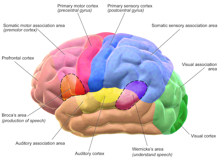
Figure showing the different regions of cortex. (credit: Blausen Medical)
This study was novel because the authors used new methods in microscopy to record the activity of cells throughout all areas of mouse cortex. The recordings were taken while mice performed a simple task: on each trial, thirsty mice were given one of two odors, and they learned that licking a spout in response to just one of the two odors yielded a water reward. You would think that this task engages olfactory areas (for smelling) as well as motor areas (for licking) and possibly decision-making and memory areas (to decide whether to lick), but you wouldn’t think that this task involves regions of cortex specialized for other functions, like vision, hearing, or touch. Indeed, silencing the activity of most regions of cortex didn’t affect the ability of the mice to correctly perform the task (i.e., lick in response to the correct odor).
The surprising discovery was that even though most of cortex isn’t required for this task, neurons all across the cortex are super active during the task. And they don’t just fire randomly—they showed activity that was linked to specific aspects of the task, like the odor turning on, the act of licking, or the delivery of water.

Figure showing neural activity (pink) across the cortex during a single trial. Activity is low before the odor turns on but is widespread during the rest of the trial. (from Allen et al., 2017)
The authors also made a bunch of cool observations comparing the activity of different types of neurons, but to me the most interesting part is what I already told you: there are parts of your brain that are not required for you do to something, but they still show very specific patterns of activity related to what you’re doing. This idea contradicts what a lot of people assume about the brain: if neurons are active at a certain time, then they must be important for whatever you’re doing at that moment. After all, why would the brain waste tons of energy by making neurons fire for no reason?
To me, that’s the big question of this paper. One answer is that it makes sense that each part of the brain would want to know what the rest of the brain is doing. Having cells across the brain that fire in similar patterns, even if most of those cells aren’t important for the ongoing behavior, could represent a form of brain-wide communication that coordinates activity across different areas.
The authors argue in favor of this idea that there are global signals being transmitted across the brain. Moreover, they identify a specific premotor region that is responsible for broadcasting information throughout the cortex. There’s a lot of work that still needs to be done, but I think this study is important in telling us that activity patterns across different areas of cortex are not only more similar than we thought, but are in fact being actively coordinated.
And more…
Ok, I’m clearly not going to be able to discuss all the other amazing research stories that came out this year, but here are a few more cool papers for you guys to check out:
“Mapping the Neural Substrates of Behavior” by Robie et al. (Kristin Branson’s lab at Janelia). In this monster paper, the authors used genetic targeting to activate over 2000 different subsets of neurons in fruit flies. They filmed the flies while these neurons were being activated, developed machine-vision technology to automatically quantify any behavioral changes, and determined which neurons and brain regions are linked to which behaviors. Whew, I’m getting tired just thinking about all that work.
“Early-Life Gene Expression in Neurons Modulates Lasting Epigenetic States” by Stroud et al. (Mike Greenberg’s lab at Harvard). The pattern of gene expression (which genes are made into protein, or “turned on”) is what defines the properties of different cell types, such as brain cells versus heart cells. As organs such as the brain develop, specific genes are permanently turned on or off in order to specify different types of cells. This study uncovered the epigenetic mechanism that mediates these permanent changes in gene expression. Moreover, the same mechanism may explain how early life experiences have long-term effects by influencing which genes get turned on or off.
“Enterochromaffin Cells Are Gut Chemosensors that Couple to Sensory Neural Pathways” by Bellono et al. (David Julius’ lab at UCSF). We’ve always known that our gut probably talks to our brain, like telling our brain we’ve been eating too much chocolate or not enough protein. This paper identifies the missing link between the gut and the nervous system: enterochromaffin cells, which are cells in the gut that are electrically excitable (just like neurons), receive chemical signals, and communicate with nerves that send information to the brain. Now you know who to blame when you feel sick after eating too much holiday candy this week.
What were your favorite neuroscience stories of the year?
Hi Anita – this is a nice review of 2017! I’d like to add to your summary of the paper from the lab of David Tank about non-spatial coding in the hippocampus: In a recent viewpoint/q&a session with nature neuroscience, a couple of neuroscientists and experts for the hippocampus (Eichenbaum, Buzsaki, Nadel, Redish, Rangananth, Lisman) were asked a set of questions more or less independently from each other, including “Do hippocampal pyramidal cells reliably encode nonspatial information?” (https://www.nature.com/articles/nn.4661) I was surprised by the amount of disagreement in their interpretation of the available body of literature, not only about this issue, but also about the other questions asked. For me (not in a hippocampus research lab), the disagreement in their opinions and the confusion which followed was maybe more informative than reading an entirely consistent, but incomplete story from a single lab, and so I would recommend it to anybody ;-)
Great recommendation, I hadn’t read that article!